
Ants, bacteria, and cells are hard to see with the naked eye but we can magnify these objects using a light microscope. As these things become smaller, we need microscopes that are more powerful and complicated – but eventually the laws of physics prevent us from seeing them. Light travels as a wave and has a certain wavelength, when we look at objects smaller than this wavelength we cannot see them. Many interesting biological molecules like DNA and proteins are smaller than the wavelength of visible light. In fact, a DNA strand’s diameter is less than a hundredth the size of light’s wavelength, making DNA invisible under a light microscope. Scientists have created a number of ways to observe these small molecules. For example, we can learn about their structure by prodding them with needles only a single atom wide. However, none of these methods are quite as simple as threading a molecule through the eye of a needle to “see” what it looks like.
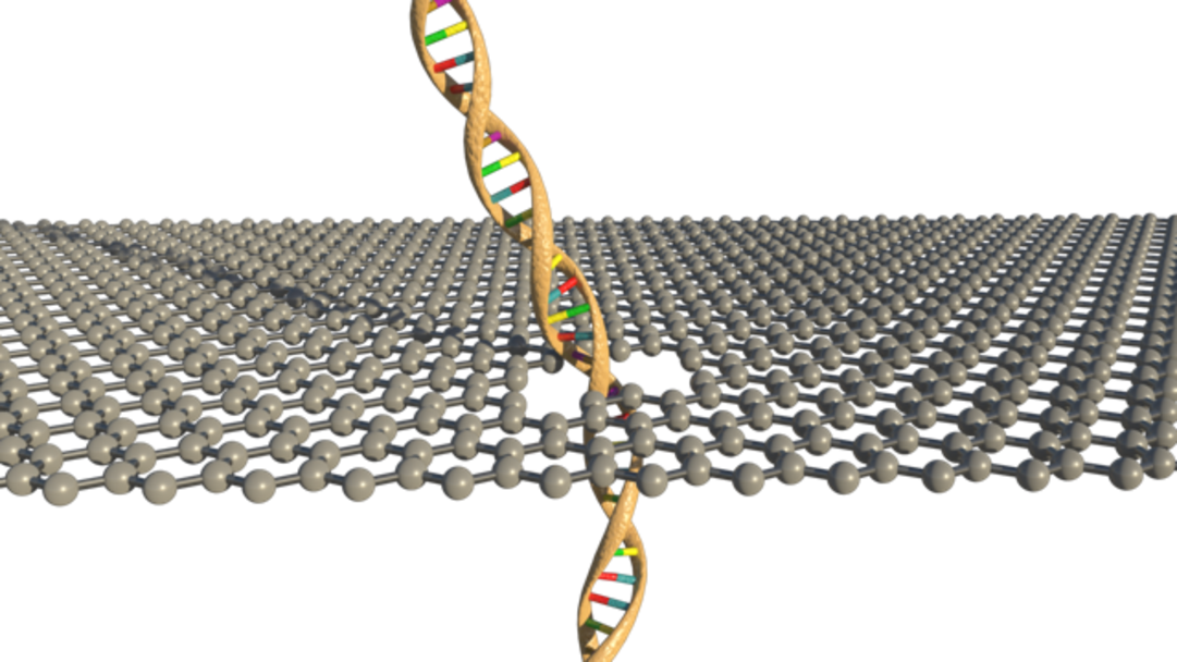
Nanopores are tubes with very small diameters of only tens of nanometres. They allow us to “see” molecules that are too small to be observed using visible light. With the help of nanopores, we can analyse biological molecules as long as they can fit through this small tube. Nanopores can be made with glass, very thin materials like graphene, or even interwoven strands of DNA which we call DNA origami. To find out information about these biological molecules we first need to fill the nanopore with ions. We can do this by applying a voltage across the tube in a salt solution, which drives ions into and through the tube, much like electrons flowing in a wire. We can then measure the flow of these ions in the nanopore, which we call the ionic current. When larger molecules like DNA or proteins enter the nanopore, it changes the number of ions that can flow through the nanopore changing the ionic current. By investigating the change in ionic current we get a molecular fingerprint containing information about the molecule’s shape and charge. This allows us to see which small molecules are present and how many there are. Using nanopores we can detect dozens of molecules including those that are indicators of health.
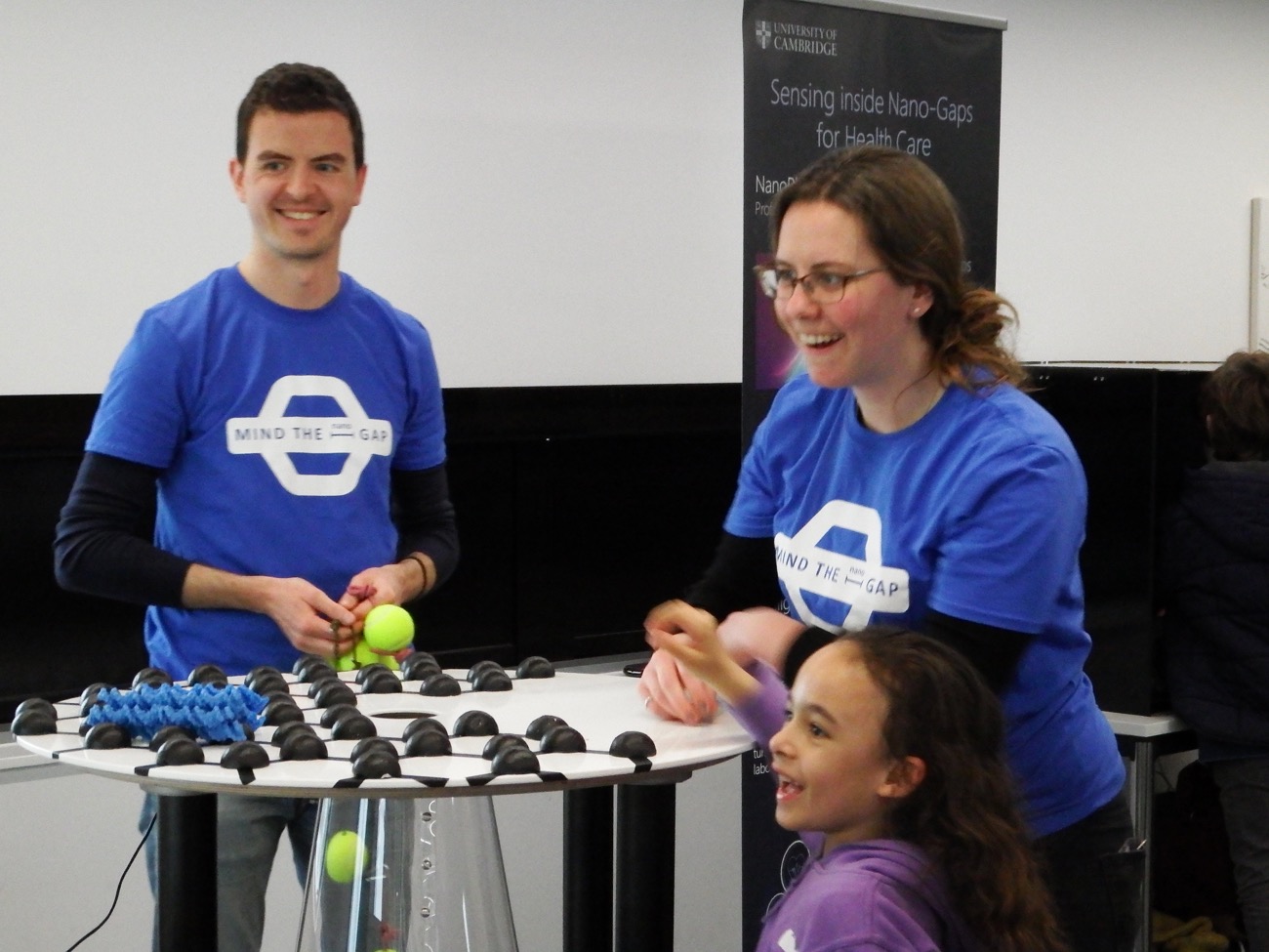
Our air tower works in a similar way, but the “current” is made of air and the molecules we want to detect are the size of tennis balls. As they fall through the “nanopore” in the centre of the tube, they block some of the air flow, which we detect as a change in pressure. Much like real nanopores, the size and duration of the pressure change allows us to find out how many balls of which size passed through the pore. Try building your own molecule and see if you can find its signature at our exhibition!
Articles on this subject:
Research Horizons- http://www.cam.ac.uk/people/ulrich-keyser
Technical Notes:
- DNA molecules and configurations in a solid-state nanopore microscope- https://www.nature.com/articles/nmat965
- DNA Origami Nanopores- https://pubs.acs.org/doi/abs/10.1021/nl204098n
- Molecule-hugging graphene nanopores- http://www.pnas.org/content/110/30/12192
- Digitally encoded DNA nanostructures for multiplexed, single-molecule protein sensing with nanopores https://www.nature.com/articles/nnano.2016.50
Groups working in this field:
![]() The Keyser Lab is a group of scientists at the Cavendish Lab, University of Cambridge, UK. Our research is focused on understanding transport processes through membranes.
The Keyser Lab is a group of scientists at the Cavendish Lab, University of Cambridge, UK. Our research is focused on understanding transport processes through membranes.
The physics of ions, macromolecules and particles in confined geometries at the single molecule/-particle level is of particular interest. We exert maximum control over all parameters in our experiments using several techniques: DNA (origami) self-assembly, optical trapping, particle tracking, fluorescence microscopy, electrophysiology, or micro-/nanofluidics, often in combination.
Our interdisciplinary team combines researchers with expertise in physics, engineering, physical chemistry, biochemistry/biology, and micro- and nanofabrication.